Thyroid and Parathyroid Blood Tests
Most patients with thyroid cancer have normal thyroid blood tests.
Most people who gain or lose weight have normal thyroid blood tests, and the thyroid gland is not the cause of the weight gain or loss. Some people who gain weight have an under functioning thyroid gland (hypothyroidism), and some people who lose weight have an over functioning thyroid gland (hyperthyroidism).
There is no need to fast for a thyroid blood test.
Thyroid function tests:
The following tests measure how your thyroid is functioning and determine if your thyroid gland is making the correct amount of thyroid hormone for you.
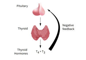
- TSH (thyroid stimulating hormone). TSH is a glycoprotein hormone. TSH is made in the pituitary gland and stimulates the thyroid to make thyroxine. The pituitary gland lies in the centre of the head near the brain.
- fT4 (free thyroxine). fT4 contains 4 iodine molecules, therefore it is called fT4. Most of T4 is bound to proteins in the blood, it is only the free portion, fT4 that is not bound to proteins in the blood, and that is the active form.
- fT3 (free triiodothyronine). fT3 contains 3 iodine molecules, therefore it is called fT3. T4 is converted to T3 in the liver and brain. Most T3 is bound to proteins in the blood, it is only the free portion, fT3 that is not bound to proteins in the blood, and that is the active form.
Hypothyroidism. High TSH and low fT4
Hyperthyroidism. Low TSH and high fT4

Notes:
fT4 and TSH act like a heater and a thermostat. When the level of T4 rises it results in the pituitary making less TSH. Conversely when TSH rises, it stimulates the thyroid to make T4.
Oestrogen in oral contraceptive tablets, and pregnancy, can increase T4 binding protein and interfere with fT4 levels.
Biotin (found in certain supplements) can interfere with thyroid function tests and should be avoided for 2 days prior to testing.
Certain medications or medical conditions may interfere with the absorption of thyroxine tablets or metabolism of thyroid hormones.
After a change in the dose of your thyroxine medication, it takes a while for the thyroid hormone levels in your blood to settle. A repeat thyroid blood test should be performed 4-6 weeks later.
Thyroid Antibody Tests
Anti-TPO (thyroid peroxidase) antibodies: Hashimoto’s thyroiditis.
TSH receptor / TSI / TR antibodies: Graves’ disease.
Anti-thyroglobulin antibodies. See section on thyroglobulin below.
Hashimoto’s Thyroiditis
Commonly we see elevated thyroid-stimulating hormone (TSH) and low thyroxine (T4) levels, coupled with increased antithyroid peroxidase (anti-TPO) antibodies.
Grave’s Disease
In Grave’s disease we see low thyroid-stimulating hormone (TSH) and high thyroxine (T4) levels, coupled with high TRAb (thyroid receptor antibody).
Thyroglobulin
Thyroglobulin is a protein made by both normal thyroid cells and cancerous thyroid cells. It contains iodine and the thyroid uses thyroglobulin to make thyroid hormones (T4 and T3).
After total thyroidectomy (removal of the thyroid), there is little, or no thyroid tissue left in the body. Therefore, the level of thyroglobulin should fall to zero or close to zero. Subsequently if the thyroglobulin level rises that suggests there is some thyroid tissue or thyroid cancer growing.
Anti-thyroglobulin antibodies can interfere with the measurement of thyroglobulin.
Thyroglobulin levels need to be interpreted in conjunction with the level of anti-thyroglobulin antibodies and the thyroid stimulating levels (TSH) in mind. A stimulated thyroglobulin level is simply the thyroglobulin level taken when the TSH is high. Conversely an unstimulated thyroglobulin level is the thyroglobulin level taken when the TSH is normal.
Calcintonin
Calcitonin is a hormone that your thyroid gland makes in the parafollicular cells of the thyroid. It helps regulate calcium levels in your blood.
Calcitonin is not routinely measured. It is elevated in medullary thyroid carcinoma-an uncommon tumour of the thyroid gland. It is measured to help establish the diagnosis of medullary thyroid carcinoma and to monitor the response to treatment of this condition.
Parathyroid Blood Tests
Parathyroid hormone (PTH) is made by the parathyroid glands. PTH is a protein that is released into the blood stream from the parathyroid glands. It plays an important role in calcium metabolism and helps control the level of calcium in the blood.
Calcium
The calcium level in the blood is tightly controlled mainly by PTH. About one third to one half of calcium in the blood is bound to albumin (a protein). Both total calcium and adjusted calcium (adjusted for the level of albumin) are measured. Adjusted calcium is also called corrected calcium. Ionised calcium represents the free, active form of calcium in the blood. In assessing patients for primary hyperparathyroidism doctors look at the adjusted calcium level.
Phosphate
Phosphate and calcium levels are closely linked and are both controlled by parathyroid hormone (PTH).
In primary hyperparathyroidism we see
- High PTH level
- High adjusted calcium level
- Low to normal phosphate level
- High 24 hour urinary calcium level
Vitamin D
Vitamin D is the sunshine vitamin and is made in the skin when exposed to ultraviolet light. Low vitamin D levels in the blood may result in an elevation of parathyroid hormone, PTH (secondary hyperparathyroidism).
Creatinine
Creatinine is a chemical waste product of creatine. The level of creatinine in the blood is a measure of kidney function. If the kidneys are not working well, then the level of creatinine rises. Chronic kidney disease may cause secondary and tertiary hyperparathyroidism.
Urea
Urea results from the breakdown of protein in the body. The level of urea in the blood is a measure of kidney function. If the kidneys are not working well, then the level of urea rises. Chronic kidney disease may cause secondary and tertiary hyperparathyroidism.
eGFR
Your kidneys filter your blood by removing waste and extra water to make urine. The estimated glomerular filtration rate (eGFR) shows how well the kidneys are filtering. A low eGFR indicates that the kidneys are not working well. Chronic kidney disease may cause secondary or and tertiary hyperparathyroidism.
Parathyroid hormone related peptide
Parathyroid hormone related peptide (PTHrP) is rarely measured. Very occasionally some tumours can make a hormone that is similar but slightly different to the usual parathyroid hormone. PTHrP is so similar to PTH that it can also cause a high calcium level. A blood test for PTH measures PTH only and does not measure PTHrP.
Urinary calcium level
Sometimes in the investigation of patients with an elevated calcium level in the blood, a 24-hour urinary calcium level is requested. This test involves collecting all your urine for 24 hours, placing it in a large container and taking that container to the lab. In primary hyperparathyroidism the 24-hour urinary calcium level is elevated. In familial hypocalciuric hypercalcaemia the 24-hour urinary calcium level is low.

